A look at the different types of pregnancy scans, courtesy of Easy Parenting Magazine
Your pregnancy test has read positive and your GP has confirmed your baby’s existence and due date. So what is the next step in antenatal care for you and your growing baby? Just what scans do you actually need, or at what stage do you need them? There are four reasons for doing ultrasound examinations of the developing baby. The first is to date the pregnancy and diagnose multiple pregnancies; the second is to screen for chromosomal abnormalities; the third is to look at how the baby is put together structurally; and the fourth is to assess foetal growth and wellbeing.
Types of scans:
Early pregnancy ultrasound scan
This scan can be performed between 8 and 15 weeks of pregnancy. However, if the scan is performed before 8 weeks, there will be little to see and you may not see the heartbeat yet. These are usually only conducted before 11/12 weeks if there is a medical concern.
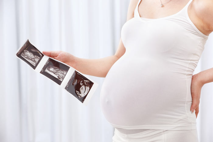
After 8 weeks you may see the pregnancy sac and the foetal pole (spinal cord beginnings) and hear your baby’s heartbeat. Most doctors advise having your first scan at 12 weeks, when the baby’s limbs, head, hands and feet will be visible. You can ask for a black and white photo of your baby at this stage.
Vaginal scans
Sometimes a vaginal scan will be required to give a clearer picture of your baby, especially if you are at an early stage of pregnancy. A vaginal transducer is lubricated and inserted into the vagina at a shallow depth. It transmits sound waves just as an abdominal transducer does. You may feel slight pressure but it is not painful and will not harm you or your baby. It may feel similar to a vaginal exam or smear test. The bladder should be empty for this type of scan.
Mid pregnancy ultrasound
This scan takes place in your second trimester at about 20 weeks. The purpose of this scan is to check that your baby is growing as expected and that everything is developing normally. It is possible to determine your baby’s gender at this point, although if they are lying in an awkward position or you have excessive stomach fat or even wind, it may not be possible to tell.
This scan is optional, although many mothers opt to have it to find out the gender, even if the main function of the scan is actually to ensure the pregnancy is going okay and not to clarify gender specifically. Some hospitals have a policy of not telling the gender to parents due to the possibility of mistakes.
Growth ultrasound scan
This scan takes place from 28 to 40 weeks in the third trimester and may only be recommended by your midwife if they feel your baby is smaller than expected for this stage of pregnancy. There are a number of other reasons why you may require a third scan, such as if you previously gave birth to a small baby, are having twins, or have complications e.g. diabetes.
What does a scan involve?
An ultrasound scan involves bouncing high frequency sound waves into your uterus. The baby’s bones will show white on the scan, and the soft tissue will look grey and speckled. The amniotic fluid surrounding baby will appear black.
The sonographer will put gel on your stomach and move a hand-held device, called a transducer, over your skin. You’ll then be able to see your baby. They will check your baby’s heartbeat and confirm if multiple pregnancies are present in the first scan.
What are the important scans?
Over the course of the pregnancy, most women will be offered two key scans. The dating scan is the first one and is done at around 14 weeks. The main reason for this scan is to date the pregnancy. It is also used to check for multiple pregnancies, see the baby’s heart beat and check for some abnormalities. The second scan is a structural scan and happens at around 20 weeks. This checks the size and development of the baby and the position of the placenta. You may be offered additional scans (discussed above) if your clinician feels they are necessary, if you have particular concerns or depending on the maternity care option you have chosen.
Are ultrasound scans safe for my baby and me?
Extensive studies have found that ultrasound is not dangerous to women or their babies. Ultrasound is merely high-frequency sound waves, inaudible to the human ear, that are transmitted through the abdomen. Ultrasound does not use radiation like X-ray tests.
Expert Advice
Dr Rachel Mackey answers some common questions about pregnancy scanning.
Q. 3D and 4D scans provide colour and black and white photographs and a DVD of the baby in the womb. Should all pregnant women go for 3D and 4D scans?
These scans are great, but they are not diagnostic. The 3 and 4 Dimensional ultrasounds are generally performed between 26 and 32 weeks of pregnancy and are not a requirement. They enhance the experience but they don’t add any medical benefits. They do not generally detect abnormalities but instead offer women and their families an opportunity to see the baby move, smile, or suck their thumbs months in advance of the birth. They are purely a matter of choice.
Q. Should a woman have a full bladder for scans?
Only in the first trimester, but scanning systems have improved so much that there is no need for this anymore.